Synapse formed by an intracellularly stained striatal spiny cell on an unlabeled dendrite. C.J. Wilson, Unpublished
LZW Compressed TIFF file 3.7MB Download (3.7MB)
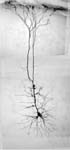
Corticostriatal neuron from the anterior cingulate cortex of the rat, stained in vivo using biocytin. From Zheng, T. and Wilson, C.J. Corticostriatal combinatorics: The implications of corticostriatal axonal arborizations. J. Neurophysiol. 87:1007-1017.
LZW Compressed TIFF file 768K Download (768K)
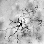
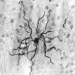
Cholinergic interneuron intracellularly stained with biocytin in 300 micron slice. From Bennett, B.D. and Wilson, C.J. Spontaneous activity of cholinergic interneurons in vitro. J. Neurosci. 19:5586-5596, 1999.
LZW Compressed TIFF file 616K Download (616K)
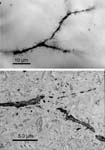
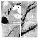
Light and electron microscopic views of the same spiny striatal cell dendrite, stained intracellularly with HRP. The light micrograph was made from a 50 micron vibratome section stained for HRP and postfixed with osmium, and embedded in resin between glass slide and coverslip. The section was cut out and glued on a block for thin sectioning. A branch of an axon from a spiny cell is seen crossing the dendrite near the branch point. C.J. Wilson (unpublished)
LZW Compressed TIFF file 2.2MB Download (2.2MB)
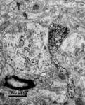
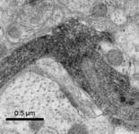
Electron micrograph of a synaptic connection between two striatal spiny neurons stained intracellularly with HRP. From Kitai, S.T. and Wilson, C.J. Intracellular labelling of neurons in mammalian brain. In: Cytochemical Techniques in Neurobiology. S. Palay and V. Chan-Palay (eds) Alan R. Liss, New York, pp.533-549, 1982.
LZW Compressed TIFF file 1.6MB Download (1.6MB)
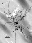
Striatal spiny neuron stained intracellularly with biocytin in vivo. From C.J. Wilson and Y. Kawaguchi, The origins of two-state spontaneous membrane potential fluctuations of neostriatal spiny neurons. J. Neurosci. 16:2397-2410, 1996.
LZW Compressed TIFF file 1.8MB Download (1.8MB)
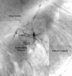
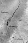
Subthalamic neuron stained intracellularly with biocytin in a 300 micron coronal slice. Stained as whole mount. Bevan, M.D. and Wilson, C.J. (unpublished).
LZW Compressed TIFF file 3.5MB Download (3.5MB)
Subthalamic neuron stained intracellularly with biocytin in a 300 micron coronal slice. Stained as whole mount. From Bevan, M.D. and Wilson, C.J. Mechanisms underlying spontaneous oscillation and rhythmic firing in rat subthalamic neurons. J. Neurosci. 19:7617-7628, 1999.
LZW Compressed TIFF file 1.6MB Download (1.6MB)
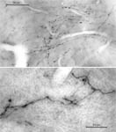
Corticostriatal axons forming varicosities in the striatum, labeled by small injections of biotinylated dextran amine in the cortex. Upper image is a light micrograph from a 50 micron section. Lower image is a high voltage electron micrograph of a 4 micron section, taken at 1 MeV on the HVEM at the University of Colorado. From Wilson, C.J. Understanding the neostriatal microcircuitry: High voltage electron microcopy. Microscopy Research and Technique 29:368-380, 1994.
LZW Compressed TIFF file 3.5MB Download (3.5MB)
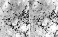
High voltage electron micrographs of enkephalin-
stained axons in the striatum, forming a stereo pair of a 4 micron section through the striatum stained for enkephalin-immunoreactivity. From Wilson, C.J. Understanding the neostriatal microcircuitry: High voltage electron microcopy. Microscopy Research and Technique 29:368-380, 1994.
LZW Compressed TIFF file 1.7MB Download (1.7MB)
High voltage electron micrographs of an intracellularly stained striatal spiny neuron (4 µm section, 500 KeV). From Wilson, C.J. Understanding the neostriatal microcircuitry: High voltage electron microcopy. Microscopy Research and Technique 29:368-380, 1994.
LZW Compressed TIFF file 4.4MB Download (4.4MB)
Spiny striatal neuron in the matrix compartment. The cell was stained intracellularly with biocytin in vivo, and 50 micron vibratome sections stained for calbindin immuno-reactivity. The calbindin-positive spiny cell somata near the cell are typical in the matrix compartment. Kawaguchi and C.J. Wilson, (unpublished)
LZW Compressed TIFF file 1.8MB Download (1.8MB)
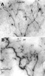
Two kinds of thalamo-striatal axonal arborizations visualized using the HVEM (4 µm section, 1MeV). The top image shows axons from the central medial/paracentral nucleus (CeM/PC); the lower image is axons from the parafascicular nucleus (PF). From Wilson, C.J. Understanding the neostriatal microcircuitry: High voltage electron microcopy. Microscopy Research and Technique 29:368-380, 1994.
LZW Compressed TIFF file 1.8MB Download (1.8MB)
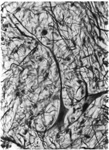
Motoneurons stained by the Bodian silver stain, which stains intermediate filaments. The darkly stained small processes are mostly axons, although some dendrites are stained well, for example the large dendrite on the neuron at the left.
LZW Compressed TIFF file 16.2MB Download (16.2MB)
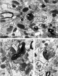
Electron micrographs of degenerating synapses in the striatum after a cortical lesion. a. A lightly myelinated axon (a) and two presynaptic boutons (b1 and b2) undergoing dark type degeneration and making synaptic contacts on the heads of dendritic spines (s). b. Degenerating (b) and normal (n) presynaptic elements making synapses on dendritic spines. c. A degenerating bouton (b) makes a synapse on a dendritic spine (s).
LZW Compressed TIFF file 13.4MB Download (13.4MB)
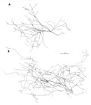
Dendritic (A) and axonal (B) arborizations of a reconstructed Striatal Cholinergic Interneuron stained by intracellular injection of HRP in vivo.
LZW Compressed TIFF file (2.6MB) Download (2.6MB)
All published images are subject to publishers' and author's copyright. All unpublished images are copyright C.J. Wilson.